Saturday, April 09, 2016

TRIOSEPHOSPHATE ISOMERASE (TPI) A DIMERIC GLYCOLYTIC ENZYME AS A MODEL OF TIM-BARREL ACTIVE-SITE STRUCTURAL AND CHEMICAL ASPECTS IN THE MONOMER LOOP REGION'S REVERSIBLE CATALYTIC REACTION.


Triosephosphate isomerase (TPI, EC 5.3.1.1) (§, ) is essential to glycolysis, catalyzes the fifth step in the glycolysis pathway the reversible conversion of dihydroxyacetone phosphate (DHAP) into glyceraldehyde-3-phosphate. TPI is a homodimer formed by two identical dimeric molecules of a single structural locus : 12p13.31. TPI has only 1 functional gene with a molecular mass of 29 kDa, that after refinement are products of a distinct single structural locus. The variant phenotype of identical subunits are expressed in both red cells and circulating lymphocytes, catalyzing the interconversion of one of the two products breakdown by reversible conversion. The TPI substrate by deprotonation the transition state reaction of dihydroxyacetone phosphate (DHAP) substrate yields one product of the glycolytic pathway, is a trend* (Kcat) that persists creating the initial complex microcompartmentation of TPI to give (G3P) glyceraldehyde-3-phosphate which seems to be the isomerase* activity, release is slower than its conversion to DHAP in normal and TPI deficient cells. TIM with its natural substrates has not been () crystalized**. TPI is a dimeric enzyme and contains 7 exons interrupted by six introns.

monomers 
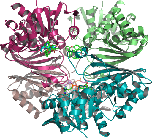
The crystallographic structure of (HsTPI) human triosephosphate isomerase PDB:1HTI is one dimer per asymmetric unit subunit 1 and subunit 2 are in the open and closed conformations in the 3-dimensional asymmetric space group P 2(1) which is specific to the Monoclinic with minimization on the entire structure in the presence of substrate analogues and its surrounding residues supporting possible regions targeted for drug design.


TPI deficiency (TPID) a disorder of glycolysis, occurring in haplotypes of specific alleles heterogeneous to clinical TPI-deficiency, with a rare homozygous deficiency the resulting genetic defect is the cause of a null variant incompatible with life by abnormally high levels of DHAP which degrades spontaneously into the toxic (MG) methylglyoxal, due to deamidation of asparagine (Asn15-71) to form aspartic and glutamic acid. Loop 6 plays a role in preventing the breakdown yield of methylglyoxal (fMG) one of the of the three products of enzyme-bound enediol(ate) phosphate, towards elimination of (fMG) inorganic phosphate. TPI deficiency is due to the common aberrant dimerization (or the dissociation into inactive monomers) of mutation TPI 1591C, encoding a Glu104-to-Asp (glutamate-to-aspartate) substitution in the TPI variant found in cases of hemolytic anemia coupled with neurodegeneration, the Glu104-to-Asp substitution is the most common disease allele inherited, when compared to wild-type TPI's three (residues from the same subunit) similar but not identical interactions between the inhibitor and catalytic residues, Glu 167 (or 165) forms a stable dimer and provides the rationale for production of structurally normal enzyme in humans, the E104D mutation, provides the amyloid-resistant structure of human triosephosphate isomerase (HsTPI). Water-protein molecules join two catalytically active monomers which is only in its dimeric form, as monomers of TIM are not functional. Within a hydrophobic catalytic pocket of the native enzymes the binding and catalysis of TPIs in hemolysates, bind to the red cell membrane. Molecular modeling using the human crystal structure of TPI was performed to determine how these mutations could affect enzyme structure and function. The Amyloid secondary structure autoepitopes antigen-driven mechanism works toward recovery of the anti-triosephosphate isomerase mutant TPI peptide** antigens. This is the scheme that allows function-enhancing stability most significantly, the catalysis for deprotonation of DHAP or vice-versa GAP substrates of the TIM-barrel relative to TPI toward turnover of two-part substrate glycolaldehyde / phosphite dianion {GA + HPO32* the transition state for this enolising enzyme substrate pieces.} Km/obsd* group of the whole GAP substrate and H95 (loop 4) is also optimal for small mutational changes in or reflects its compatibility with amino acid residues which stabilizes the enediolate intermediate (GA/HPO) activity from change in the products scheme (a proton transfer mechanism) DHAP/G3P or interconversion of these intermediates.

dhap-g3p

Closed (activated for catalysis) of optimal WT (TPI) molecular modeling PDB 1HTI_B using the human crystal structure of TPI human triosephosphate isomerase (HsTPI) conformation 1hti_b, calculated to the incidence residue Water-protein molecules and the protein cage that interacts within a hydrophobic catalytic pocket isolated and examined which coded for human triose-phosphate isomerase. [EC: 5.3.1.1]….


The active flexible site loop must open before product release, unliganded in trypanosomal Tb-TIM glycerol phosphate ester to liganded Glu167 in the catalytic cycle and the enzymes substrate transition state between open and closed to protect the substrate for the turnover of DHAP and G3P (GAP) the natural substrates, and inhibiting the formation of a toxic by-product in the absence of this equilibration reactions between dihydroxyacetone phosphate and glyceraldehyde 3-phosphate (G3P) enzymes by mutations that impair biosynthesis transforming competent cells, in the presence of an auxotrophic effect with these differences generated for an inability of the host organism to synthesize an essential compound during glycolysis in Tb-TIM. Trypanosomal-TIM is a glycolytic enzyme essential for the parasite survival that causes Chagas* disease, in this study G. bellum from the genus related Geraniaceae and its phenolic compound are leads which generates an unstable epimer of an enzyme Geranin A-containing changes resulting from ligand adducts in the active site to capture in addition a source of frustration that becomes more favourable. Glycolaldehyde (GA) the simplest sugar-related molecules uptake of a proton by Glu167 preserves the small effect for inhibition by PGA (transition-state analog) relative to substrate, G3P produces a triosephosphate isomerase with wild-type activity, loop 6 adopts the "closed" desolvated  (+) conformation to facilitate completion of catalysis by the formation of the › Michaelis-Menten complex (on the ‹ micros-ms › time scale) utilization yields further corrected calculations with corresponding (slower Kcat) motional rates* Km. Increase's are discussed in the context of the significance (Enzyme kinetics\Kcat) and may be estimated where the 'single-substrate' is locked in a protein cage probably  because of an active reaction site (loop 6) movement to the transition state for deprotonation; which are the on-average opened (substrate binding and release) and closed (activated for catalysis) of both monomers optimal WT (wild type) TIM conformations. Lys-12 ‹ is expected to interact with both centers, where the enediol intermediate along with the catalytic glutamate base and histidine-95 the catalytic electrophile stabalizes the reversible reaction intermediate that polarizes the substrate DHAP in the Michaelis complex. Interconversion spans the C-terminal end of the eight β-strands. For catalysis to occur likley a low pKa value transition from DHAP - for the enolase reaction enzyme enhancement 'relative to the nonenzymatic reaction - (Bound PGH - phosphoglycolohydroxamate mimics the (closed form) negative polarization () charge••, while PGA (2-phosphoglycolate) the positively charged residues in the two active conformation sites.) is similar for the two conformers' in the closed conformation, on ligand binding interacting with the reactive end's (β) the deprotonated substrate-bound structures to be protonated by a single-base (Glu-165) proton transfer^ mechanism.

philo

Structure of human triose phosphate isomerase at the positions of introns in homologous TPI genes from a number of phylogenetically diverse species. The introns motif are identified as calculated in phylogeny.
Phylogenetic trees constructed on the basis of sequence comparisons for triosephosphate isomerases analysis, TIM sequences were constructed based phylogeny with similarity, to those adopting the same structural fold of interest from different species for the taxonomic groups and the K13M mutations involvement in the human triosephosphate isomerase gene family...


Interactions in the loop regions combine the effects of His95 and Lys13 for Glu165 (loop 4, 1, and 6) the three crucial catalytic residues in triose phosphate isomerase, all form the enediol intermediate necessary for the interconversion reaction catalyzed by TIM resulting in the natural substrates G3P formation. The introns motif are identified as calculated in phylogenic motifs. Poorly conserved residues as targets for specific•• drug design are expected when compared to (TPI) Triosephosphate isomerase (•). Catalytic residues of the phylogenetic relationship pathways obtained by sequence based methods of specific key amino acids can than be calculated to the incidence residues and other TIMs which may influence the (human) HsTPI equilibrium.


Friday, November 20, 2015

Non-Phosphorylating And Phosphorylating Oxidoreductase Glyceraldehyde-3-Phosphate Dehydrogenase As Part Of A Structure-Based Design In Glycolysis As The Glycolytic Protein G3PD.

Glyceraldehyde-3-phosphate dehydrogenase (EC 1.2.1.12) GAPDH1/G3PD, is located in band 12p13.31; related to both glycolysis2 and gluconeogenesis-pathways. G3PD catalyzes reversible oxidative phosphorylation of inorganic phosphate and nicotinamide3 adenine dinucleotide (NAD)4 converting in glycolysis the glycolytic protein GAPDH5 in which adenosine-triphosphate (ATP)6 is generated when phosphoglycerate kinase (PGK)7 is produced in the GAPDH8-catalyzed reaction. These intermediate metabolites (aldolase9, triose-phosphate10-isomerase (TPI)11) catalyze the Glycolysis reactions, in the sequence of the ten enzyme-catalyzed Embden12-Meyerhof13 reactions in the metabolic pathway. Converting phosphoglycerate mutase 1 (PGM)14 catalyzing the internal steps by 2,3-BPG15 phosphatase to form by converting D-glyceraldehyde 3-phosphate (G3P)16 into 1,3-bisphosphoglycerate (1,3-BPG)17 from its role as 3-Phosphoglyceric acid (3PG) in glycolysis as the glycolytic protein GAPDH18 that catalyzes the first step (G3P19 into 1,3-BPG) of the pathway. Plant20 cells contain several reactions of photosynthesis21 participating in glycolysis and the Calvin-Benson22 cycle signaling pathways in plants (cytosolic-GAPC23 (Arabidopsis thaliana)24 functions in plant25 cells.) its final byproduct is also another Glyceraldehyde-3-P. GAPDH is a band 326 protein that associates with the cytoplasmic27 face of human erythrocyte28 (RBC)29 membranes. The cytoplasmic GAPDH exists primarily as a tetrameric30 isoform, 4 identical 37 kDa31 subunits. By subcellular translocation GAPDH32 participates in nuclear events [In nuclear membrane the vesicular*33 tubular cluster fractions34 (VTCs)35 - anterograde transport or retrograde36 membrane transport complexes37 between the intermediates, these are the Golgi38 complex and the endoplasmic reticulum (ER)39, in the nucleus a function is lost in disease* that exploits this process.], this a change to a non-cytosolic40 localization due to the signal transduction pathways (considering Lm41GAPG L.42 mexicana43-like functions.) involved in s-nitrosylase44 activity that mediates, governed by the equilibrium between four cysteine residues (nitrosylation45 and denitrosylation reactions)46, inhibition of GAPDH nuclear translocation, as a basis47 for its multifunctional48 activities relating to the extraglycolytic functions of GAPDH. Nuclear GAPDH49 promotes glucose metabolism to sustain50 cellular ATP51 levels, or potentially by inhibiting targets52 of p30053/CBP such as p5354 dependent phosphorylation. Nitric oxide synthase or neuronal NOS ( involved in cellular and human intracellular55 nuclei events56, in addition to the cytoplasm) could generate nitric oxide57 (NO). GAPDH has four cysteine58 residues which are associated with S-nitrosylation59-yielding NOS60-GAPDH which “recruited” its glycolysis subunit61 from the three63 molecular axes translocation roles (S-thiolation64, S-nitrosylation or aggregated65 enzymes (Cys-15266 and nearby 15667 converted into a 'cross-linked68 soluble' states)), and (SNO69-GAPDH) nitrosylated S-nitrosoglutathione70 (GSNO)71 the active site cysteine residue in GAPDH at its Cys 15072 residue that binds to Siah1 (seven in absentia homolog 1) acquisition and the translocation of GAPDH into the nucleus, and denitrosylation using a combination of approaches, including G3P73 . And NADPH may play a role in (VTC) vesicle74 function. The complex would function in the apoptosis cascade75 by its molecules translocation, this may76 depend on lysine 22777 in the human GAPDH78-Siah79 interaction to another intracellular position80 induced by apoptotic81 stimuli, augments p30082/CREB binding protein (CBP)-associated83 acetylation of nuclear proteins. 'Engineering the cofactor (GAPDH-(Lys) K160R84-K227A) availability prevents85 activation of p300/CBP that interferes with GAPDH-Siah1 binding'86-prevents the ternary (GAPDH-Siah1) complex associations translocation; that CGP-346687 can reduce independently with both cofactors88. Dysregulation of protein S-nitrosylation (S-nitrosocysteine89 - 247) by lipopolysaccharide (LPS) is associated with pathological90 conditions which contributes to disease phenotype, where GAPDH protects ribosomal protein RP91-L13a92 from degradation, L13a93 and GAPDH94 forms a functional GAIT95 complex. One of the functions of GAPDH proteins role in glycolysis96 in relation to DNA97 synthesis is nuclear accumulation associated by the NAD98(+)-dependent s-nitrosylation99 and denitrosylation01 reactions both of these isforms are distinct02 parallel to the uracil DNA glycosylase (UDG)03 gene in mitochondria04 and in the nucleus is N-terminally processed is the 37-kDa subunit05 of the (GAPDH)06 glyceraldehyde-3-phosphate dehydrogenase protein. This enzyme is an example of moonlighting protein which is validated and replaced07 by alternative reference genes that link (in their nuclear forms) on the multifunctional08 properties of the enzyme GAPDH09 known as a key enzyme in glycolysis that contributes to a number of diverse cellular functions unrelated00 to glycolysis001 depending upon its subcellular location. GAPDH is a key enzyme in glycolysis the most commonly used expression is as a housekeeping002 gene.


GAPDH-Siah1Cytotoxic stimuli [1a.] or Programmed cell death, via nitric oxide generation, lead to the binding of GAPDH from its usual tetrameric form to a dimeric form, to the protein Siah1 [1.] intracellular G-3-P [2.] substrate [3.] protects GAPDH from S-nitrosylation [4.]. The GAPDH-Siah interaction depends on lysine 227 [5.], in human GAPDH that interacts with a large groove [6.] of the Siah1 dimer, that connects the GAPDH dimer to PGK in the cytoplasm. figure7The S-nitrosylation [7.,8.] abolishes catalytic activity and confers upon GAPDH the ability to bind to Siah [9.]. (GAPDH) is physiologically nitrosylated at its Cys 150 residue. GAPDH (SNO-GAPDH) [10.] binds to Siah1 [11.] by forming a protein complex. In the nucleus [12.] GAPDH is acetylated at Lys 160 [13.] and binds to the protein acetyltransferase p300/CBP. Under these conditions siah-1 formed a complex with GAPDH (PDB:4O63) and localized in the nucleus of Müller cells [14.]. GAPDH mutants [15.] that cannot bind Siah1 prevents translocation [16.] to the nucleus to elicit neurotoxicity [17.] and cell apoptosis.
[1a.] 16492755, 8769851003 [1.]16391220, [2.]19542219, 22534308, 3350006004, 19937139, [3.]22847419, [4.]15951807, [5.]20601085, [6.]16510976, 20392205005, [7.,8.]22817468006, 16505364007, [9.]16633896, [10.]16574384, [11.]20972425, [12.]19607794, [13.]18552833, [14.]19940145, [15.]23027902008, [16.]24362262, [17.]16492755.


H placental GAPDHAnalysis of CGP-3466 Docking (NAD) to Human Placental GAPDH which decreases the synthesis of pro-apoptotic proteins is N-terminally PMID:10677844, processed to which a Rossmann NAD(P) binding fold as seen in figure 1 is a C-terminal domain as seen on this page, PMID:10617673, 26022259, 16510976 ...The structure is also used to build a model of the complex between GAPDH and the E3 ubiquitin ligase Siah1. (Purple Ribbon-1U8F_Q Figure 1.)


(Figure 3.) Glycolysis and GlyconeogenesisIn the GAPDH-catalyzed reaction these intermediate metabolites (aldolase, triose-phosphate-isomerase Glycolysis and Glyconeogenesis (TPI)) catalyze the Glycolysis reactions, in the sequence of the ten enzyme-catalyzed Embden-Meyerhof reactions in the  metabolic pathway. Converting phosphoglycerate mutase 1 (PGM) catalyzing the internal steps by 2,3-BPG phosphatase to form by converting D-glyceraldehyde 3-phosphate g3p(G3P) into 1,3-bisphosphoglycerate (1,3-BPG) from its role as 3-Phosphoglyceric acid (3PG) in glycolysis as the glycolytic protein GAPDH that catalyzes the first step (G3P into 1,3-BPG) of the pathway.


(Figure 4.) GAPDH homotetramerGAPDH homotetramer was studied as represented an assembly of repeating spherical units that harbored a distinct birefringent crystal structure to the optic axis for the p polarization, also (r axis) discernible via transmission electron microscopy. of the relative amount of soluble monomeric GAPDH to G3P in the binding pocket of the NAD(+)-binding site residue located at the active site linked to GAPDH in Figures 5 and 6. PMID:10407144009, 25086035.


g3pAnother model building studie indicates that a structure obtained where glyceraldehyde 3-phosphate PDB:3CMC_Q binds in the P(s) pocket of the natural substrate G3P phosphorylating GAPDH (PDB:1U8F_Q) at the catalytic cysteine residue site. To define the conditions suitable for affinity for the cosubstrate, the isolation and accumulation of the intermediate metabolites per G3P monomer found in Figure 8 of the equivalent Glc-3-P structure in the binding pocket of the NAD(+)-binding site residue located at the active site linked to GAPDH. PMID:19542219, 22534308


APO/STPCorrectly known binding sites on ((GAPD/NAD)) structures, polar spheres of the binding catalytic pocket that corresponds to G3P (glyceraldehyde 3-phosphate) aligned to the holographical structure nonbounded spheres (salmon color), these apoenzymes together with the cofactor(s) Cys 151, 152 which corresponds as below the Ps pocket of G3P, on the Green ribbon required for cofactor activity. Together with eliminated crystallographic waters and other possible spheres, these are at least one atom of a amino acid residue in contact with at least one alpha sphere of one binding pocket on the holo protein NAD structure 1U8F_Q needed to align holo and apo structures included in this data set with G3P (PDB:3CMC_Q) was tested only on holo structure (NAD), obtained via Pea Green spheres aligned to 1U8F_Q ribbons/ligand structure which provide structural recognition insights into the biological 1U8F-Q assembly this includes 29 asymmetric units of its dimeric form, along the tetrameric 1U8F biological forms axis. PMID:9461340010


siah1-pdb:4i7d_g3pd-pdb:1u8f(Figure 8.) These are the results without the liquid chromatography coupled mass spectrometer, that are known 3D products by two-dimensional sequence analyses with the STRAP alignment tools data sets and which may have any effect on the functions of further analysis involved in more ordered results than this study attempts to show, of the analysis that may be included are identified separated into multiple gradients here in these paired graphs. Therefore in the present work to uncover the exact coincidence of 1U8F_R and 4I7D_C, the 3D coordinates of GAPDH (PDB:1U8F_Q) to the protein Siah1 4I7D were not presenting when subjected to STRAP  alignment this apparent discrepancy (Figure 1.) was partially resolved by a (Figure 7) rendering from a more reactive native GAPDH_R homotetramer model.


References:

Sunday, June 14, 2015

CHANGES IN GLUTATHIONE AND GLUTATHIONE REDUCTASE POSITIONING GLUTATHIONE-S-TRANSFERASE AS A FUNCTION OF CELL CONCENTRATION WITH ENZYME ACTIVITIES FOUND TO INFLUENCE BEHAVIOR.

Glutathione reductase (GSR, GR) locus in the chromosomal region 8p21.1, (EC 1.8.1.7)-(§, ) is a protein-S-glutathionylation, as a (human) Mitochondrial localization of hGSR and its associated enzymes cellular thiol/disulfides S-Glutathione reductase (GSR) which is the importance of significance in reversible thiol modifications which  regenerates reduced glutathione (GSH) and GSSG to the reduced form found in the obvious structural properties of glutathione reductase. The redox regulating enzymes relationship with TTase (thioltransferase) activity with the ratio of the activities of G3PD, as the mechanism (of cellular repair) 'differs' (gssg-g6pg) according to the type of reducing glutathionylated mixed disulfide, including protein-S-S-glutathione (PSSG), GSR reduces (PSSG) modified by thiolation to a normal level in human lens epithelial (HLE) cells. This may have implications in stress- and aging-related pathologies in astrocytes and granule cells, demonstrated by comparable mitochondria/cytosolic concentrations of its thiol proteins, where a mitochondrial leader sequence (cDNA) is present in the gene structure of human GSR and may be the Cytoplasmic Isoform (derivative or inhibitor formed) of  mitochondrial dysfunction that contains the catalytic cysteine revealing a possible therapeutic strategy/target, also indicating transiently accumulated inhibitor proteins modified by thiolation (cysteine catalytic subunits) compounds that inhibit these (re)activation processes (hGSR) with its structure-based prosthetic group (FAD) cofactor is common because of the levels of cysteine available; are mitochondria/cytosolic concentrations that the Glutathione reductases reversible thiol modifications which catalyzes the reduction of GSSG to GSH the natural GR substrate is dependent on the NADPH:GS-SG ratio.
PDB Id: 3DK9 Cys58 and Cys63 represent the enzyme's results seen as the reductive (GSH) Cys-58 and oxidative (GSSG) Cys-63 is the relationship of these two enzymes, His467' is seen to interact with Cys63 more optimally and Cys-58 produces the second GSH intermediate molecule of the reaction is the reduced glutathione-to-oxidized glutathione ratio (GSH/GS-SG) when compared to the substrate free form correlated with (FAD) the flavin compounds, flow from NADPH to the substrate GSSG via flavin. The reducing equivalents needed for regeneration of GSH are provided by NADPH. The enzyme has affinity for flavin adenine dinucleotide (FAD) the prosthetic group of GR, and maintains high levels of reduced glutathione  (Cytoplasmic Isoform: Produced by alternative initiation of isoform Mitochondrial homodimer, derivative or inhibitor formed from the GSR Pyridine, dimerisation domain.) in the cytosol. Glutathione reductase (GR) plays a key role in maintaining either a thiol group or a nonprotein sulfhydryl group (NPS) form of GSH, and potential for thioredoxin and glutathione systems, as thioredoxin dose not require GSH and GR for catalytic activity. Glutathione reductase (GR) utilizes NADPH produced by G6PDH (glucose-6-phosphate dehydrogenase) enzyme activities, and enzyme glutathione reductase (GR) represents the erythrocyte glutathione-reducing system (GRS), of the GSH pathway to oxidation and inactivation in the activity of GSH peroxidase and GSH reductase. Expression of the regulatory subunit of gamma-glutamylcysteine synthetase/ligase (GCL) catalyzes the first and rate-limiting step in the production of the cellular (GSH) glutathione. Dietary riboflavin (Vitamin B2) intake produces its active essential coenzyme flavin forms, riboflavin mononucleotide (FMN) and flavin adenine dinucleotide (FAD) of glutathione reductase (GR), or the GR activity correlated with red-cell flavin compounds.When both GSSG and NADP(+) substrates and products are present, glutathione reductase (GR) is a enzyme required for the conversion in the presence and absence of flavin adenine dinucleotide (FAD), glutathione reductase (GR) is an obligatory FAD-containing homodimer. GSSG via glutathione reductase (GR) regenerates reduced glutathione which is essential for antioxidant defense. The flavoenzyme glutathione reductase (GR) reduces 'oxidized glutathione' (GSSG) back to GSH, also involving glutamate-cysteine ligase and modulatory (GCL)-can be upregulated ∉ as the cellular GSH system, indicating short-term and heritable tolerance of exposure to oxidative stress from/via numerous reporting ∈ mechanisms. NADPH is used by glutathione reductase for the reduction of oxidized glutathione (glutathione disulphide) GSSG to GSH-dependent peroxide metabolism. 4-Hydroxynonenal (HNE) is one of the major end products of lipid peroxidation which may lead to enhanced action of  the (GSR) oxygen radical, glutathione S-transferases (GSTs) are specifically suited to the detoxification and removal of 4-HNE (∋ or ∝) from cells which may provide a basis for selective cellular and/or subcellular distribution of mitochondrial and cytosolic to individual detoxifying gene inducer activities of glutathione reductase (GR), the cellular (GSH) glutathione. It was evident the enzyme glutathione reductase (GR) represents the erythrocyte glutathione-reducing system (GRS), of the GSH pathway to oxidation and the (∉ or ∝) inhibition constant for reversible inactivation in the activity of glutathione related antioxidant enzymes. And GSH reductase may be one of the factors that remained in focus that suggests its effects on the antioxidant system related to glutathione synthesis (GCL), degradation, and functions.

Biological Xenobiotics, Extracts, Applications of note In the presence of Glutathione reductase.:

Schisandrin (Schisandra chinensis), used in traditional Chinese medicine. PMID:21328628
Transketolase (TK) and transaldolase (TA)
Melatonin PMID:15571523, 19475625
Blackberry (Rubus sp.) cultivars, The 'Hull Thornless',  PMID:11087537
Glutathione dehydrogenase (ascorbate)-[dehydroascorbate reductase (DHAR), and glutathione reductase (GR). This enzyme participates in the glutathione metabolism the active metabolite of vitamin D3 increases glutathione levels.] PMID:11087537, 23770363
3H-1,2-dithiole-3-thione nutraceutical D3T potently induces the cellular GSH system, Anethole trithione is a drug used in the treatment of dry mouth, the Anethole trithione isomer is related to anethole (anise camphor) used as a flavoring substance. PMID:17206382*, 19408115,     19176875*, 15896789, 18408143*, Glutathione reductase
16946404*
Cassia fistula used in herbal medicine. PMID:19088944
Sanguinarine is extracted from some plants, including bloodroot and Mexican prickly poppy (Argemone mexicana) where argimone oil causes Epidemic dropsy. PMID:11260782
Vitamin E, PMID: 15672860
Tocotrienols are natural compounds members of the vitamin E family found in select vegetable oils are an essential nutrient for the body. PMID:21845802
Pyrrolizidine alkaloids are produced by plants as a defense mechanism against insect herbivores consumption of PAs is known as pyrrolizidine alkaloidosis. PMID:20144959
Apple extract (AE) PMID:20401791
Lipoic Acid an organic compound, forming a disulfide bond, available as a dietary supplement PMID:15246746, 21073761
Carnitine PMID:15246746, 10581232
Vitamin D upregulated expression of GCLC and GR. PMID:23770363
Vitamin D3_ PMID:12416023
Vitamin E_ PMID:10459841, 8360018, 18296478, 21845802, 15490422, 16885600, 7062348, 20729758, 21086752
Shidagonglao roots Mahonia fortunei (十大功劳 shi da gong lao) species contains the alkaloid berberine PMID:199382 18
Coenzyme Q10 (CoQ10) PMID:16621054
Trigonella foenum graecum seed powder (TSP) PMID:15026271
Boschniakia rossica, a ̱̱̱Traditional Chinese medicine. PMID:19352025
Aegle marmelos commonly known as bael is a species of tree. PMID:18830880
Scoparia dulcis A medicinal plant, dulcis. PMID:21905284
Fenugreek (Trigonella foenum-graecum)  is used as a herb. PMID:15026271
L-arginine (L-Arg) semiessential supplementation common natural amino acid. PMID:16038634
Hypericum perforatum (St. John's Wort) PMID:18754092
Urtica dioica often called common nettle PMID:12834006
Usnea longissima, a medicinal lichen. PMID:16169175
Capparis decidua, a fruting tree also used in folk medicine and herbalism. PMID:22272107
Indole-3-carbinol found at relatively high levels in cruciferous vegetables such as broccoli
PMID:9512722, 14512388
Ascorbate Vitamin C. PMID:14512388
Sulforaphane It is obtained from cruciferous vegetables such as broccoli. PMID:12628444, 18607771*, 22303412
Andrographis paniculata, may shorten the duration and lessen the symptoms of common cold. PMID:11507728
Vitamin B-1 (thiamine) PMID:1132146, 10450194, 21308351*, 11514662*, 1270885
Vitamin B2 (riboflavin) PMID: 5822598, 5550591, 1201246, 5794396, 237845, 3677785, 3582603, 12194936, 2721660, 1261528, 5721130, 14608016, 4400882, 7883462, 844948, 7337797, 5881,12641409, 4393763, 3497609, 16883966...(№ 1244, OMIM.138300)
Vitamin B-6 (Pyridoxine) PMID:2721660, 3582603, 10450194, 15490422, 1270885, 7417521, 7337797, 7814235
Vitamin B9 (Folic acid)  PMID: 844947, 1270885
Aspartate transaminase (AST) or glutamic oxaloacetic transaminase (GOT) catalyzes the interconversion of aspartate an important enzyme in amino acid metabolism. PMID:1132146, 10450194, 1253408
β-Carotene is a strongly colored red-orange pigment abundant in plants and fruits. PMID:19957244
3-Hydroxykynurenine (3OHKyn) a metabolite of tryptophan. PMID:11273669
Ajoene ((E,Z)-4,5,9-trithiadodeca-1,6,11-triene 9-oxide), a garlic-derived natural compound. PMID:9986706 PDB: 1BWC
Propolis a product made by bees. PMID:19394397
Resveratrol produced naturally by several plants PMID:12797471
 No CiTO relationships defined:
 http://vixra.org/abs/1506.0104
 http://www.citeulike.org/user/emissrto/article/13645622

Tuesday, March 03, 2015

Thioredoxin reductase: Selenotetrapeptide sequences with specificity for thioredoxin and glutathione systems

  Thioredoxin reductase (EC 1.6.4.5) TXNRD1 (Alternate Symbols: GRIM-12, TR, TRXR) chromosomal position 12q23.3-q24.1 (§, ) is a homodimeric selenocysteine-containing enzyme. Secys a selenocysteine residue is an essential TR isozyme component, located near the C-terminus region [cysteine (Cys)-497,Secys-498] of the intracellular, redox cellular environments center in the catalytically active enzyme site, Gly-499 is the actual C-terminal amino acid. In their N-terminal sequences Cys-59, Cys-64 links the thiol/disulfide oxidoreductase dependent pathway reductases from there to the flexible C-terminal part (Secys) of the other sub cellular subunit by which Selenocystine is efficiently reduced and induce RNR (Ribonucleotide reductase) for replication and repair, where Trx reductase (TR) or oxidized GSH (GSSG) reductase further supply electrons for RNR. The protein reversibly modulates specific signal transduction cascades, to regulate multiple downstream intracellular redox-sensitive proteins that links NADPH and thiol-dependent processes which catalyzes NADPH-dependent reduction in the presence of the redox protein-Trx and thioredoxin reductase (TR) maintain cysteine residues in numerous proteins in the reduced state. There are three TXNRD selenoproteins  5-prime end variants essential for mammals, one V3 (TXNRD1) encodes an N-terminal glutaredoxin (GRX) these variants code for thioredoxin glutathione reductases (TGR). V3 associates with and triggers formation of Filopodia (cytoplasmic filaments) can guide actin in migrating cells, the emerging protrusions of cell membrane restructuring involved is in 'deglutathionylation values" for mitochondrial and cytosolic thioredoxin reductase (TR) domains. Characterization of the TR native Thioredoxin and glutathione systems (TGR) suggests that the lifecycle of E. granulosus and Schistosoma mansoni a phylum of Platyhelmintha, involves the TXNRD1_v3 isoform containing a fused (Grx) glutaredoxin domain which is abolished by deglutathionylation' targeted to either mitochondria or the nucleus in the reduction of glutathionylated substrates, in leishmaniasis (disease) glutathione reductase system (TGR) is replaced by the trypanothione reductase (TcTR) system in mammalian cells, essential as these TR3 are significant as a recognized drug target of these (TcTR) human protozoan parasites. Cytosolic TR1, mitochondrial - TR3 and TrxR2 (locus 22q11.21) where TrxR1 and TrxR2 are consdered as the respective cytosolic 1w1e MITOCHONDRIAL cytoplasmicand mitochondrial thioredoxin reductases, plus the thioredoxin glutathione reductases-TGR systems most likely can reduce (Trx) by fusion of the TR and an N-terminal glutaredoxin domains. As a pyridine nucleotide disulfide oxidoreductase of the oxidized GSH and GSSG (selenodiglutathione) reductase TGR structures enzyme stability, are linked to the previously characterized two thioredoxin reductases cytosolic TR1 and TR3, and one mitochondrial variant. Selenols are key metabolites at mammalian TXNRD1's active (SeCys 498) site. Thioredoxin undergoes NADPH-dependent reduction (NTRs) and reduce oxidized cysteine groups on mitochondrial TXNRD1 proteins similar to the cytosolic enzyme, from the FAD binding domain where the active cystines and the NADPH binding domain are contained, plus an interface domain (ID) of the C-terminal interface homologous to glutathione reductase identifies a mechanism of p53 mediated cell death regulation involving (TrxR) enzymes of redox homeostasis reactions to overcome the oxidative stress generating reactive oxygen species (ROS) on a complex combination of decreased apoptosis to prevent permanent cell damage and cell death that tumor cells use to evade the redox-sensitive signaling factors, or resistance to therapy. End products of lipid-peroxidation, 4-HNE-(4-Hydroxynonenal) can induce oxidative stress, other isoforms are more water-soluble adducts detoxifying such a buildup,  peroxidation might be limiting their (selenoproteins) proper expression. Thioredoxin reductase (TrxR) is the homodimeric flavoenzyme that catalyzes reduction of thioredoxin disulfide (Trx) one of the major redox control systems, involving a second interaction between NAD(P)H and/or (quinone reductase) NQO1 via the FAD-containing enzyme (TR), thioredoxin reductase forms an oxidoreductase system. TrxRs are able to reduce a number of substrate proteins other than Trx.


3qfbThe 3' UTR of selenocysteine-containing genes have a common stem-loop structure, the sec insertion sequence (selenocystine-SECIS, PDB: 2ZZ0), that is necessary for the recognition of a catalytically active Sec codon rather in the values for mitochondrial and cytosolic thioredoxins reductase (TR) domains. The Sec residue is protonated at a different pka than in comparison to that of Cysteine. Cys59-Cys64 two cysteines pair also was oxidized in the N-terminal FAD domain essential for thioredoxin-reducing activity, and the need for Sec-498 (PDB: 2J3N) to be in complex with the FAD and NADP(+) during catalysis to the N-terminal active site cysteine residues Cys59-Cys64 and from there to the C-terminal part of the other subunit which have selenotetrapeptide sequences from the other module (PDB: 2J3N). Secys498 forms, (Human PDB 3QFB,) can both be identified at active site of the enzyme Gly-499 of the subunits active Cys-497-TRXR1 (the TR1 structure PDB: 3QFB) are the mechanism(s) for the incorporation of Se into TrxRs as the amino acid selenocysteine (Sec), as well as for delivery to a variety of secondary substrates or TRX (PDB: 3QFB) in nuclei provide means to quantify glutathione (GSH) (PDB: 3H8Q) conditions of the active GRX functonally and structurally analogus to TGR (selenodiglutathione) reductase. These two were modeled parts of TGR were linked to V3 (_TXNRD1) encodes an N-terminal inter-specific glutaredoxin (PDB: 1JHB).3qfb-3h8q From the FAD binding domain-(PDB: 1ZKQ ) active cystines and the NADPH binding domain where they are contained, plus an interface domain (ID) of the C-terminal ID in complex with its substrate thioredoxin (Trx-PDB: 1TRX, TXNRD1-3QFB) bringing Cys32 in Trx1 close to Cys497 in 3H8Q to quantify glutathione (GSH) that helped in characterizing  what was separately modeled as the Thioredoxin reductase (TXNRD1) domain which are consdered as the respective cytosolic and mitochondrial thioredoxin reductases units with a model obeying standard geometry that is conceivable of human thioredoxin reductase 3's structure  glutaredoxin domain 3H8Q  in complex with the FAD and NADP(H), when replaced by the TcTR (PDB: 2W0H) trypanothione/trypanothione reductase system involves a phylum of Platyhelmintha, where a glutathione (GSH) isoform containing a fused (Grx) glutaredoxin domain  (PDB: 1JHB) is essential for the parasite survival.  The intricate substrate specificities for the thioredoxin (Trx) system which consists of native Trx and the respective cytosolic  mitochondrial thioredoxin reductase (TrxR) enzymes are likely to be of central importance to these observations as a determinant of TrxR function in general, each (the thioredoxin reductase/thioredoxin pathway) can reduce a number of different types of substrates or cross-reactive-bound enzyme fractions as active with thioredoxin.


[1.] Selenium yeast: seleno yeast PMID: 16857846
[2.] Sulforaphane From Broccoli PMID: 16377050, 12742546, 20204301, 12949356, 19595745, 17150329, 15740016, 12663510, 15998110, 17300148
[3.] Chlorella vulgaris: corresponding to a chloroplast NADPH-dependent thioredoxin reductase gene (NTR-C), in Chlorella PMID: 18029787
[4.] Scutellarin:  It can be found in Scutellaria barbata and S. lateriflora. PMID: 15131321
[5.] Curcumin (TURMERIC plant of the ginger family): PMID: 21782934, 20160040, ~15879598
[6.] Experiments in E. huxleyi genus phytoplankton PMID: 20032866
[7.] Gambogic Acid pigment of gambooge resin from tree species Garcinia gummi-gutta. PMID: 24407164
[8.] Shikonin an antioxidant (no longer approved for use,: targets the Sec residue [13.] in TrxR1 to inhibit its physiological function. see: (Methane-) methylseleninic acid (MSA)) obtained from the extracts of  plant [9.] Lithospermum erythrorhizon. PMID: 24583460
[10.] Black tea extract (BTE) theaflavin (TF) PMID: 19059456
[11.] Green tea extract-epigallocatechin-3-gallate (EGCG) PMID: 19020731
[12.] Eicosatetraenoic acid, (Mortierella Alpina Oil) Arachidonic acid (AA) all-cis-5,8,11,14-eicosatetraenoic acid, 5-Hydroxyicosatetraenoic_acid_and_5-oxo-eicosatetraenoic_acid PMID: 15123685
[13.] Juglone: In the food industry known as C.I. Natural Brown 7 and C.I. 75500. (DTNB assay, a synthetic approach for Cys and Sec residues.) PMID: 21172426, 11170645, 18382651 ... a 5,5'-[dithiobis Pyritinol: analogue, Sulbutiamine]
[14.] The antioxidant ubiquinol-10 (Q10) PMID: 12435734
[15.] Rottlerin, conductance potassium channel (BKCa++) opener, source the Kamala tree. PMID: 17581112
[16.] Ajoene a chemical compound available from garlic. PMID: 9986706
      No CiTO relationships defined
http://vixra.org/abs/1502.0252
http://www.citeulike.org/user/emissrto/article/13530556

Monday, November 24, 2014

Catalase, the antioxidant heme enzyme one of three subgroups related to catalase deficiency in humans modulating the normal catalase reaction dependent on NADPH-binding catalases for function.

Catalase (CAT) is converted by decomposition and intracellular localization relationships of the main cellular antioxidant enzyme system like superoxide dismutase (SOD), peroxiredoxins (Prdx), and glutathione peroxidase (GPX) are peroxisomal matrix enzymes in the cytoplasm, translocated to the peroxisomes to catalyze hydrogen peroxide H2O2 which is decomposed to oxygen and water, locus: 11p13 (§, ). Unlike catalase, the objective of this communication, SOD which prevents the formation of Hydroxyl radicals - (HRGT) determined from constant of O2.- dismutation, and generation of reversibly inactive (CAT)-compound II, Panax ginseng could induce both transcription factors. Catalase is  composed of four identical subunits each of the subunits binds one heme-containing active site, and produces two catalase compounds HPI and HPII (PDB: 1p80) is flipped 180 degrees » with respect to the orientation of the heme related to the « root mean square to the structure of catalase, (Mutation Location) from peroxisomal catalases inactive state in compound II NADP+(H) binding pockets inverted remains similar to the structure of the wild type (Val111, PDB:1A4E, KatG) orientation on the heme proximal (PDB: 1GGK) side, inactivate catalase can be prevented by melatonin. Catalase (CAT; EC 1.11.1.6) a  free radical scavenging enzyme (FRSE) is a scavenger of H2O2. Protoporphyrin - (ZnPPIX) (PDB: 1H6N), from a heme group of the 'heme-pathway, which forms catalase,' is a scavenger of antioxidant (HO-1-HMOX1) heme oxygenase, involving ROS. Catalase is part of the enzymatic defense system constituting the primary defense against ROS, zinc protoporphyrin IX (ZnPPIX) is an inhibitor of (HO-1) heme oxygenase. Catalase protects the cell from oxidative damage by the accumulation of cellular reactive oxygen species (ROS) generation systems, those peroxisomal enzymes that breaks down hydrogen peroxide after H(2)O(2) exposure, and thereby mitigates* (some contradictory* results) the toxic effects of hydrogen peroxide. In the process (The typical hydroperoxidases (CAT) known as Compound I) of the substrate of catalase, NADP+ (an inactive state, compound II) is replaced by another molecule of NADP(H) to provide protection of catalase against inactivation by (H2O2) hydrogen peroxide. Erythrocyte  [Human erythrocyte catalase (HEC), The NADPH-binding sites were empty - PDB: 1F4J, 1QQW] and plasma indices (enzymatic-antioxidants) initially implies the thiobarbituric acid-reacting substances (TBARS) based on reaction with hydroxyl radicals (OH) can release thiobarbituric acid, TBAR inhibition measures malondialdehyde (MDA - impact of coenzyme Q10) correlated (with MPO-myeloperoxidase activity -generating ROS) as co-variable, by which mulberry leaf polysaccharide (MLPII) via the decomposition of (certain) MDA, products of lipid peroxidation (LPO) were reduced. Comparisons were to specific activities of catalase (SNP) single nucleotide polymorphisms (CAT-C-262 (rs1001179) the low-risk allele) of genetic variants in both, promoter a common C/T polymorphism (262-C/T), and in nine - exonic - regions and its boundaries, occur frequently associated distally in genomic mutations, similar to those of normal catalase demonstrating changes in catalase protein level targeted to the peroxisomal matrix. The 262-C/T CAT low-risk allele is hypothetically related to the lower risk variant allele CAT Tyr308 G to A point mutation ineducable in the Japanese acatalasemia allele. The common C/T polymorphism can be targeted by dietary and/or pharmacological antioxidants, and the endogenous antioxidant defense enzymes concentration can prevent cellular lipid (LPO) peroxidative reactions occurring. Catalase is a homotetramer complex of 4 identical monofunctional subunits. Catalase is located at the peroxisome of human cells associated with several (PBDs)-peroxisomal biogenesis disorders commonly caused by mutations in the PEX genes, peroxisomal targeting signal 1 (PTS1) protein affecting in peroxisomal biogenesis, the monomeric to homotetrameric transition in the forms of peroxisome biogenesis disorder. PBDs also include Acatalasemia the only disease known to be caused by the (CAT) gene. In human catalase, the antioxidant heme enzyme, is localized in the cytoplasm to the peroxisome, nucleus, or linked with mitochondria which in most cells lack catalase (Peroxisomes do not contain DNA), its mitochondrial fraction (microperoxisome), a secondary phenomena shows physiological decline, aging and age-related reactions in mitochondrial function and disfunction. NADPH is required for the prevention of forming an inactive state of the enzyme. Antioxidative defence mechanisms, capacity and redox cycle enzyme activities increasing with Tc treatment Tinospora cordifolia (Tc), T and B cells and antibody. Both RBCs and plasma were measured on parameters of oxidative stress. Syzygium cumini aqueous leaves extract (ASc) was able to remove oxidant species in a hyperglycemic state generated in red blood cells RBC-CAT levels. Catalase alone is unable to prevent in a hyperglycemic state. Macrophages recruit other types of immune cells such as lymphocytes white blood cells (WBCs).  Catalase is dependent on the family of NADPH-binding catalases for function, the prevention and reversal of inactivation by its toxic substrate (H2O2) hydrogen peroxide. Amyloid-beta binds catalase and inhibits (H2O2) hydrogen peroxide, a reactive oxygen species, breakdown through efficient dismutation, and malonaldelhyde (MDA) determined in plasma, as well as another member of the oxidoreductase family, myeloperoxidase (MPO (EC 1.11.1.7)) converting H(2)O(2), the reducing equivalents produces (HOCl) hypochlorous acid a mechanism of cell-mediated antimicrobial immune defense for monofunctional catalases one of three subgroups related to catalase deficiency in humans, in micro-organisms manganese-containing catalases ('large catalases') determining in part the bifunctional activity of (KatG, PDB:1X7U) represented by bifunctional (heme) catalase-peroxidase based Bacterial-resistance mechanisms. Peroxiredoxins (Prxs, EC 1.11.1.21), bifunctional catalase-peroxidases (KatGs) two organelle systems are antioxidant enzymes of the peroxiredoxin family that oxidize and reduce H(2)O(2) hydrogen peroxide thereby modulating the catalase reaction, KatGs are not found in plants and animals. Trx (thioredoxin) a redox-regulating protein also controls the antioxidant enzyme activity of the main cellular antioxidant enzymes (AOE) superoxide dismutase (SOD) and catalase.


The function of NADPH bound to Catalase.
catalaseThe cytosine to thymidine transition of nucleotide-262 (-262C>T) Computer analysis indicated that the two variants bound promoter the Ile  (-262 C/T) and (B) Ile-262 in the 5'-flanking region carrying the T allele best captured and characterized the generation of the hydroxyl radical site in (PDB: 1DGB), (CAT) -[GLU] 330C>T transition, is known also as -262C>T. The 'T allele in comparison to the C allele' is a common C/T polymorphism frequency in the promoter region association was observed between genotypes for locus11p13 risk alleles acatalasemia mutation Asp (37C>T in exon 9) was hypothetically related to the lower risk Japanese acatalasemia allele Tyr308 a single G to A (see: rs7947841  to evaluate the link to rs769214) point mutation ineducable or near exon 9 (TC, CC, TT) of the CAT gene to which variant changes in the promoter region C/T-262 polymorphism are more closely related to CAT T/C at codon 389 in exon 9 (rs769217) polymorphism did not differ significantly from those of healthy controls in both promoter (-262 C/T) and in exonic (ASP-389 C/T) regions of the catalase (CAT). catalase Tyr 370 resolves the 25 A-long (hydrogen peroxide) channel a constriction or narrowing of the channel leading to the heme cavity ('Parameters) situated in the entrance channel to a heme protoporphyrin (ZnPPIX) (PDB: 1H6N) from a heme group, capable of heme biosynthesis' in a wide range of organisms convert it into into heme b, protoporphyrin IX-heme. Two channels lead close to the distal side.  A third channel reaching the heme proximal side Tyr 370, Ile-262 is proposed as a the 'PDB: 1DGB - variant with a substituted residue in the ASP 178 to the (Met) D181E variant PDB 1p80'.  These differences include the structure of the variant protein Val111Ala (Saccharomyces cerevisiae) related supports the existence of the 'Heme and NADP(H) binding pockets'. The omission of a 20-residue  PDB: 1F4J, (1QQW) segment corresponds to the N-terminal (blue) of catalase from human erythrocytes (HEC), or in a C-terminal (red) domain organized with an extra flavodoxin-like fold topology may provide with weak coordination the N- or C-terminal, that allows scrutiny of the origins (topology) in this report of what would otherwise remain speculative or determined with further verification.

 Biological Xenobiotic Extracts Applications of note In the presence of Catalase:

green tea polyphenol (-)-epigallocatechin-3-gallate (EGCG)
Yamamoto T, Lewis J, Wataha J, Dickinson D, Singh B, Bollag WB, Ueta E, OsakiT, Athar M, Schuster G, Hsu S. Roles of catalase and hydrogen peroxide in greentea polyphenol-induced chemopreventive effects. J Pharmacol Exp Ther. 2004Jan;308(1):317-23. Epub 2003 Oct 20. PubMed PMID: 14569057.Furukawa A, Oikawa S, Murata M, Hiraku Y, Kawanishi S. (-)-Epigallocatechingallate causes oxidative damage to isolated and cellular DNA. Biochem Pharmacol.2003 Nov 1;66(9):1769-78. PubMed PMID: 14563487.*
Trigonella (Fenugreek)
Mohammad S, Taha A, Bamezai RN, Basir SF, Baquer NZ. Lower doses of vanadatein combination with trigonella restore altered carbohydrate metabolism andantioxidant status in alloxan-diabetic rats. Clin Chim Acta. 2004Apr;342(1-2):105-14. Erratum in: Clin Chim Acta. 2010 Aug 5;411(15-16):1158.Mohamad, Sameer [corrected to Mohammad, Sameer]. PubMed PMID: 15026271.
Aegle marmelos
Khan TH, Sultana S. Antioxidant and hepatoprotective potential of Aeglemarmelos Correa. against CCl4-induced oxidative stress and early tumor events. JEnzyme Inhib Med Chem. 2009 Apr;24(2):320-7. doi: 10.1080/14756360802167754 .PubMed PMID: 18830880.
Centella asiatica
Flora SJ, Gupta R. Beneficial effects of Centella asiatica aqueous extractagainst arsenic-induced oxidative stress and essential metal status in rats.Phytother Res. 2007 Oct;21(10):980-8. PubMed PMID: 17600859.
Daidzein
Mishra P, Kar A, Kale RK. Prevention of chemically induced mammarytumorigenesis by daidzein in pre-pubertal rats: the role of peroxidative damageand antioxidative enzymes. Mol Cell Biochem. 2009 May;325(1-2):149-57. doi:10.1007/s11010-009-0029-1. Epub 2009 Feb 13. PubMed PMID: 19214712.
Capparis
Yadav P, Sarkar S, Bhatnagar D. Action of capparis decidua againstalloxan-induced oxidative stress and diabetes in rat tissues. Pharmacol Res. 1997Sep;36(3):221-8. PubMed PMID: 9367667.
Retinal
 Kannan R, Jin M, Gamulescu MA, Hinton DR. Ceramide-induced apoptosis: role ofcatalase and hepatocyte growth factor. Free Radic Biol Med. 2004 Jul15;37(2):166-75. PubMed PMID: 15203188.
Retinol
Cemek M, Caksen H, Bayiroğlu F, Cemek F, Dede S. Oxidative stress andenzymic-non-enzymic antioxidant responses in children with acute pneumonia. CellBiochem Funct. 2006 May-Jun;24(3):269-73. PubMed PMID: 16634091.
Diallyl disulfide (Allicin)
Kalayarasan S, Prabhu PN, Sriram N, Manikandan R, Arumugam M, Sudhandiran G.Diallyl sulfide enhances antioxidants and inhibits inflammation through theactivation of Nrf2 against gentamicin-induced nephrotoxicity in Wistar rats. EurJ Pharmacol. 2009 Mar 15;606(1-3):162-71. doi: 10.1016/j.ejphar.2008.12.055. Epub2009 Jan 19. PubMed PMID: 19374873.
Leucas aspera (Catechin, EGCG)
Kripa KG, Chamundeeswari D, Thanka J, Uma Maheswara Reddy C. Modulation ofinflammatory markers by the ethanolic extract of Leucas aspera in adjuvantarthritis. J Ethnopharmacol. 2011 Apr 12;134(3):1024-7. doi:10.1016/j.jep.2011.01.010. Epub 2011 Jan 18. PubMed PMID: 21251972.
Urtica dioica (nettle suppliment)Ozen T, Korkmaz H. Modulatory effect of Urtica dioica L. (Urticaceae) leaf
extract on biotransformation enzyme systems, antioxidant enzymes, lactatedehydrogenase and lipid peroxidation in mice. Phytomedicine. 2003;10(5):405-15.PubMed PMID: 12834006.
Justicia adhatoda
Singh RP, Padmavathi B, Rao AR. Modulatory influence of Adhatoda vesica(Justicia adhatoda) leaf extract on the enzymes of xenobiotic metabolism,antioxidant status and lipid peroxidation in mice. Mol Cell Biochem. 2000Oct;213(1-2):99-109. PubMed PMID: 11129964.
Phyllanthus niruri L. (Euphorbiaceae) (P. niruri)
Bhattacharjee R, Sil PC. Protein isolate from the herb, Phyllanthus niruri L.(Euphorbiaceae), plays hepatoprotective role against carbon tetrachloride inducedliver damage via its antioxidant properties. Food Chem Toxicol. 2007May;45(5):817-26. Epub 2006 Nov 11. PubMed PMID: 17175085.
Tinospora cordifolia
Sharma V, Pandey D. Protective Role of Tinospora cordifolia againstLead-induced Hepatotoxicity. Toxicol Int. 2010 Jan;17(1):12-7. doi:10.4103/0971-6580.68343. PubMed PMID: 21042467; PubMed Central PMCID: PMC2964743.
Aher V, Kumar Wahi A. Biotechnological Approach to Evaluate theImmunomodulatory Activity of Ethanolic Extract of Tinospora cordifolia Stem(Mango Plant Climber). Iran J Pharm Res. 2012 Summer;11(3):863-72. PubMed PMID:24250513; PubMed Central PMCID: PMC3813135.
coenzyme Q10
Lee BJ, Lin YC, Huang YC, Ko YW, Hsia S, Lin PT. The relationship betweencoenzyme Q10, oxidative stress, and antioxidant enzymes activities and coronaryartery disease. ScientificWorldJournal. 2012;2012:792756. doi:10.1100/2012/792756. Epub 2012 May 3. PubMed PMID: 22645453; PubMed CentralPMCID: PMC3356738.
Dietary carotenoid-rich pequi oil
Miranda-Vilela AL, Akimoto AK, Alves PC, Pereira LC, Gonçalves CA,Klautau-Guimarães MN, Grisolia CK. Dietary carotenoid-rich pequi oil reducesplasma lipid peroxidation and DNA damage in runners and evidence for anassociation with MnSOD genetic variant -Val9Ala. Genet Mol Res. 2009 Dec15;8(4):1481-95. doi: 10.4238/vol8-4gmr684. PubMed PMID: 20082261.
Tinospora cordifolia  (Mango Plant Climber) extract from Tinospora known as Tinofend Aher V, Kumar Wahi A. Biotechnological Approach to Evaluate theImmunomodulatory Activity of Ethanolic Extract of Tinospora cordifolia Stem(Mango Plant Climber). Iran J Pharm Res. 2012 Summer;11(3):863-72. PubMed PMID:24250513; PubMed Central PMCID: PMC3813135.
 mulberry leaf polysaccharide (MLPII)
Ren C, Zhang Y, Cui W, Lu G, Wang Y, Gao H, Huang L, Mu Z. A polysaccharideextract of mulberry leaf ameliorates hepatic glucose metabolism and insulinsignaling in rats with type 2 diabetes induced by high fat-diet andstreptozotocin. Int J Biol Macromol. 2014 Oct 11. pii: S0141-8130(14)00674-6.doi: 10.1016/j.ijbiomac.2014.09.060. [Epub ahead of print] PubMed PMID: 25316427.
five widely studied medicinal plants (Protandim)
Nelson SK, Bose SK, Grunwald GK, Myhill P, McCord JM. The induction of humansuperoxide dismutase and catalase in vivo: a fundamentally new approach toantioxidant therapy. Free Radic Biol Med. 2006 Jan 15;40(2):341-7. PubMed PMID:16413416.
melatonin
Mayo JC, Tan DX, Sainz RM, Lopez-Burillo S, Reiter RJ. Oxidative damage tocatalase induced by peroxyl radicals: functional protection by melatonin andother antioxidants. Free Radic Res. 2003 May;37(5):543-53. PubMed PMID: 12797476.
Protective effect of harmaline
Kim DH, Jang YY, Han ES, Lee CS. Protective effect of harmaline and harmalolagainst dopamine- and 6-hydroxydopamine-induced oxidative damage of brainmitochondria and synaptosomes, and viability loss of PC12 cells. Eur J Neurosci.2001 May;13(10):1861-72. PubMed PMID: 11403679.
horseradish peroxidase (HRP)
Shen L, Hu N. Heme protein films with polyamidoamine dendrimer: directelectrochemistry and electrocatalysis. Biochim Biophys Acta. 2004 Jan30;1608(1):23-33. PubMed PMID: 14741582.
Selegiline (--)Deprenyl
Kitani K, Minami C, Isobe K, Maehara K, Kanai S, Ivy GO, Carrillo MC. Why(--)deprenyl prolongs survivals of experimental animals: increase of anti-oxidantenzymes in brain and other body tissues as well as mobilization of varioushumoral factors may lead to systemic anti-aging effects. Mech Ageing Dev. 2002Apr 30;123(8):1087-100. Review. PubMed PMID: 12044958.
Rhodiola rosea
Bayliak MM, Lushchak VI. The golden root, Rhodiola rosea, prolongs lifespanbut decreases oxidative stress resistance in yeast Saccharomyces cerevisiae.Phytomedicine. 2011 Nov 15;18(14):1262-8. doi: 10.1016/j.phymed.2011.06.010. Epub2011 Jul 30. PubMed PMID: 21802922.
Carnitine
Kiziltunc A, Coğalgil S, Cerrahoğlu L. Carnitine and antioxidants levels inpatients with rheumatoid arthritis. Scand J Rheumatol. 1998;27(6):441-5. PubMedPMID: 9855215.
 Syzygium cumini
 De Bona KS, Bellé LP, Sari MH, Thomé G, Schetinger MR, Morsch VM, Boligon A,
Athayde ML, Pigatto AS, Moretto MB. Syzygium cumini extract decrease adenosine
deaminase, 5'nucleotidase activities and oxidative damage in platelets of
diabetic patients. Cell Physiol Biochem. 2010;26(4-5):729-38. doi:
10.1159/000322340. Epub 2010 Oct 29. PubMed PMID: 21063110.